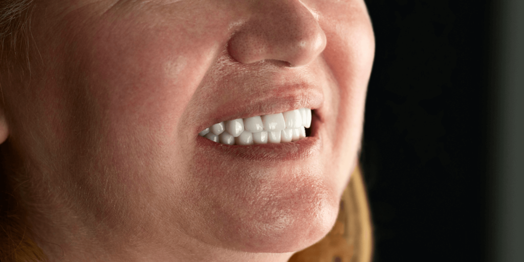
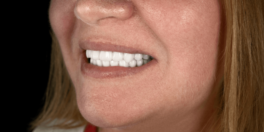
Specializes in: surgery, implant placement, and dental prosthetics
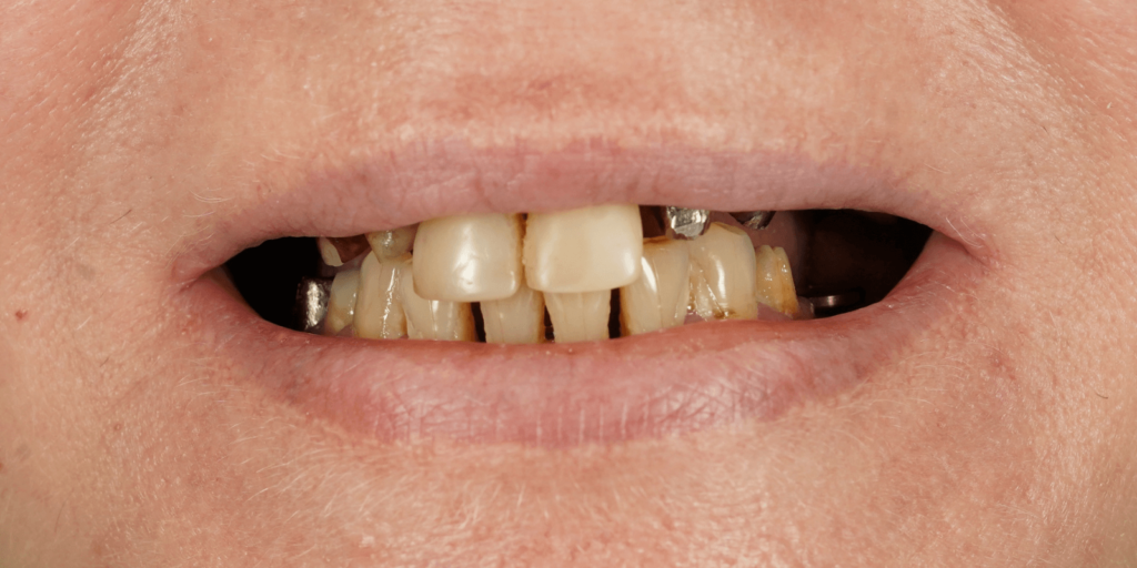
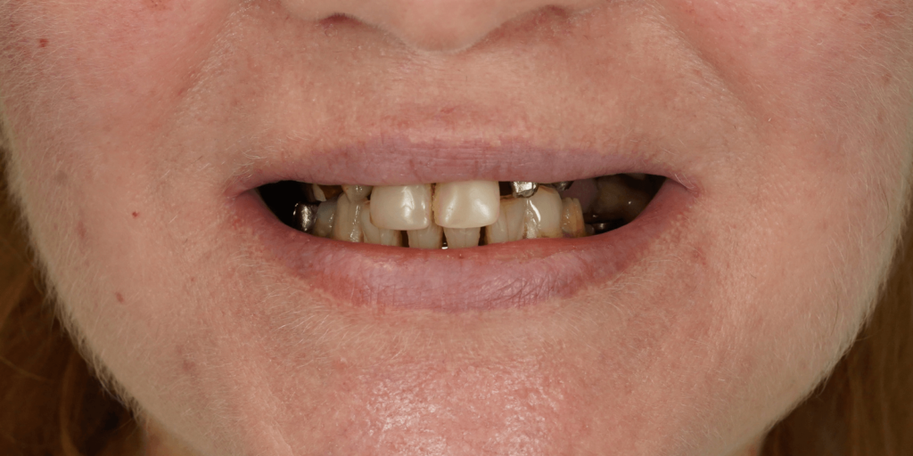
The patient came to our clinic with complaints about an aesthetic defect of the dentition, tooth mobility, and jaw pain. During the examination, the doctor found:
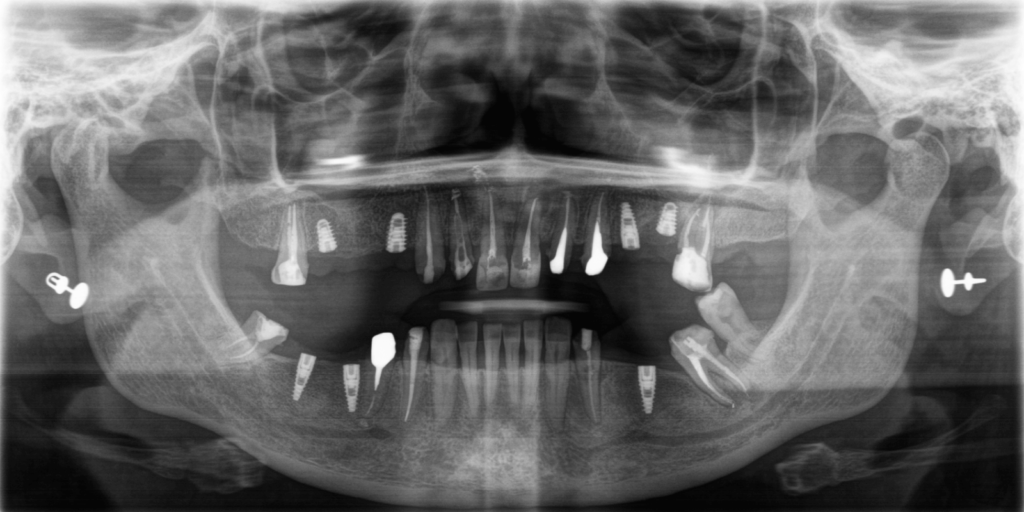
In the upper jaw: 2 bridge-like prostheses, 4 missing teeth, a dental crown on tooth 2.6, and carious cavities in two anterior teeth.
In the lower jaw: 2 bridge-like prostheses and 4 missing teeth. Tooth 3.8 was mobile and in contact with the antagonist in the upper jaw.
All prosthetic structures were metal-ceramic and needed replacement for several reasons:
The condition of the teeth under the structures was unsatisfactory; the structures were worn and had poor aesthetics, and some abutment teeth needed to be extracted.
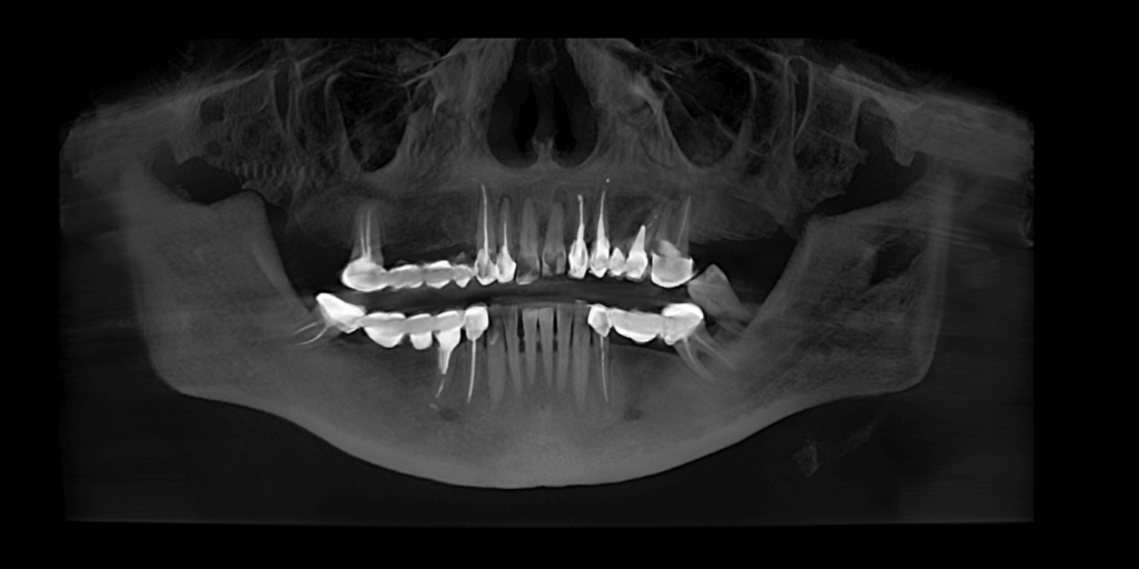
In addition, during the CT assessment, the doctor found granulomas and cystogranulomas around the roots of almost all abutment teeth under the bridge structures.
Therefore, our team, which in this case included a periodontist, an endodontist, a general dentist, a prosthodontist, and an implant surgeon, developed the following treatment plan:
Remove all prosthetic structures, perform tests, and identify the teeth that could be preserved.
Perform endodontic treatment of the teeth that could be preserved.
Extract teeth 2.5 and 3.8 due to a high degree of mobility and advanced periodontitis.
Place 4 dental implants in the upper jaw and 3 in the lower jaw.
During prosthetic treatment, take into account the need to raise the bite.
Perform prosthetic treatment using a mock-up model.
Use temporary dental crowns and bridges to improve the patient’s quality of life during the rehabilitation period and to approve the future shape and size of the teeth.
Fabricate permanent prosthetic restorations from multilayer zirconium dioxide.
Treatment consisted of the following stages:
STAGE 1
Examination. Treatment plan
A CT scan was performed, based on which the team of doctors created a treatment plan while following the patient’s main request: "the teeth need to be fixed quickly and beautifully."
STAGE 2
Professional oral hygiene
We begin every treatment with professional hygiene to reduce inflammation and clean the oral cavity of pathogenic bacteria. This is especially important before implant placement.
STAGE 3
Secondary endodontic treatment
Over several visits, the doctor performed secondary root canal treatment on 12 teeth and treated lesions beyond the root apex (cysts).
STAGE 4
Surgery
Tooth extractions and implant placement were performed in a single-stage procedure. The surgery was carried out under local anesthesia. After that, the doctor provided recommendations and prescribed medication therapy.
Our driver then took the patient home comfortably and safely.
STAGE 5
Scanning. Fabrication and try-in of the mock-up model
The doctor scanned the jaws, transferred the data to software, modeled the mock-up, then printed it and transferred it to the patient’s oral cavity.
STAGE 6
Preparation. Placement of temporary crowns
After 3 days, the mock-up model was processed and the teeth were prepared for permanent prosthetics. The prepared teeth were scanned again by the doctor, after which temporary crowns were placed.
STAGE 7
Fixation of permanent crowns
One week later, the doctor fixed the permanent crowns, as well as bridges supported by implants and natural teeth.
SUMMARY
Treatment lasted about 1.5 months.
Doctors involved in the treatment: Lidiia Bulyhina, Maksym Makarenko, Denys Kaliberda.