A patient came to us complaining of mobile bridges on the upper and lower jaws, an inability to chew properly, and inflamed, bleeding gums.
We proposed the following treatment plan:
- perform professional hygiene of the oral cavity;
- carry out root canal retreatment of the lower-jaw abutment teeth that could be preserved;
- extract the teeth that could not be restored;
- place six Straumann BLX implants in the upper jaw using the All-on-6 protocol;
- place six Straumann BLX implants in the lower jaw;
- carry out prosthetics of both jaws.
The treatment consisted of the following stages:
1 ЕТАП
Examination. Treatment plan
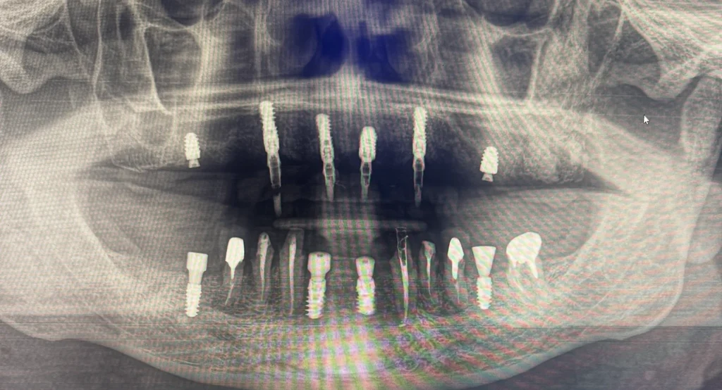
After a clinical examination and a CT scan we identified a number of problems: the bridge on the upper jaw had lost its cement, which caused one of the abutment teeth it was attached to to break; teeth 11, 12, 21 and 22 were significantly destroyed by a long-standing carious process; the anterior teeth of the lower jaw — 41, 42, 31 and 32 — had become mobile against a background of pronounced gum inflammation, and one of the lower chewing teeth was completely destroyed. Some of the lower-jaw teeth could be preserved provided they received endodontic treatment. We also took the medical history into account: the patient had chronic maxillary sinusitis on both sides, so we planned the position of the upper-jaw implants with this condition in mind.
2 ЕТАП
Professional oral hygiene
We started the treatment with professional hygiene of the oral cavity.
3 ЕТАП
Endodontic retreatment
Our endodontist retreated the root canals of four lower-jaw abutment teeth — premolars 34, 35, 44 and 45 — which we decided to keep as support for the future bridges.
4 ЕТАП
Surgical stage
The next day we performed the surgery. In the upper jaw we removed the teeth that could not be preserved and placed six Straumann BLX implants using the All-on-6 protocol. In the lower jaw we removed the mobile anterior teeth (41, 42, 31, 32) and the destroyed chewing tooth, placed six implants in the anterior and chewing sections, performed bone grafting where necessary, fitted gum formers and carried out a digital scan. Given the chronic maxillary sinusitis, the patient received antibiotic therapy before and after the surgery.
5 ЕТАП
Temporary restorations
Three days after the surgery we fixed a temporary fixed prosthesis on the upper jaw — the implants received early loading straight away. On the lower jaw we placed a temporary restoration supported by the patient’s own teeth, while the implants integrated under the gum formers without loading.
6 ЕТАП
Permanent prosthetics
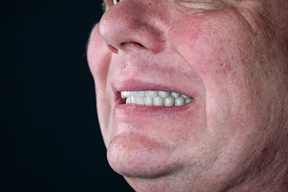
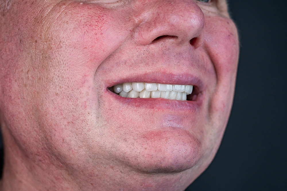
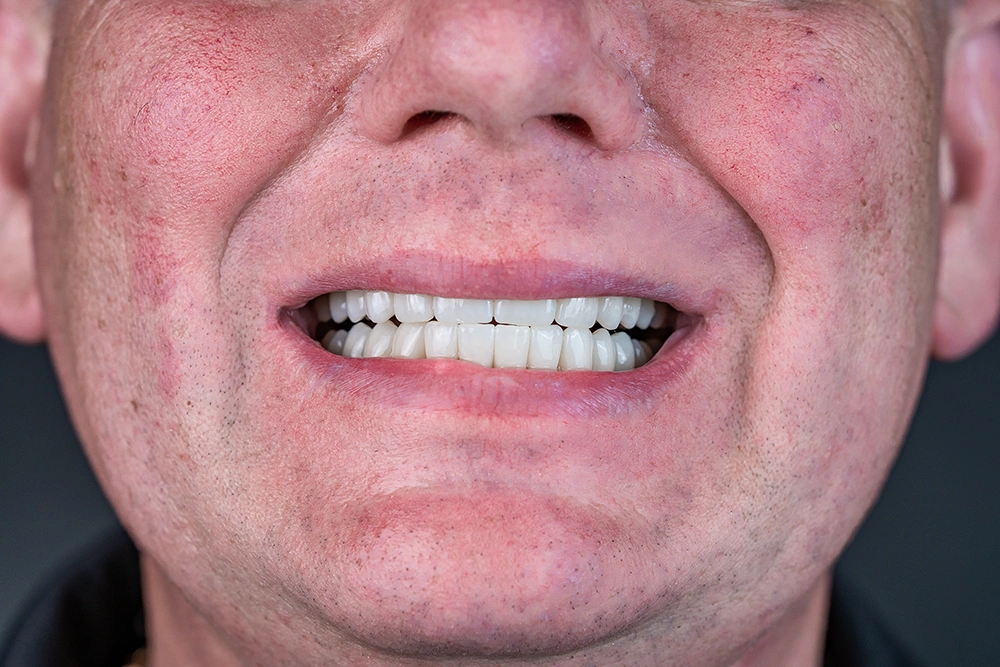
Two and a half months later the patient came for a check-up. The implants had integrated successfully, so we removed the temporary restorations, performed another digital scan and made the permanent prostheses. On the upper jaw we fixed a zirconium dioxide prosthesis on a titanium bar. On the lower jaw we placed bridges supported by the implants and the patient’s own premolars (34, 35, 44, 45). The shades were selected together with the patient: A2 for the upper jaw and A3 for the lower — so the smile looks natural.
Treatment outcome
We fully restored chewing function, the stability of the restorations and the aesthetics of the smile. We scheduled a follow-up X-ray examination in three months to assess the condition of the implants and the healing process. The patient is happy with the result and has returned to a comfortable life without pain or restrictions while eating.